The humerus is the long bone in the arm that run from the shoulder to the elbow. It plays a essential purpose in the motility and stability of the upper limb. Understanding the humerus bone markings is indispensable for aesculapian pro, anatomists, and students of human anatomy. These markings function as attachment points for muscles, ligament, and tendon, and they provide worthful information about the os's office and construction.
Anatomy of the Humerus
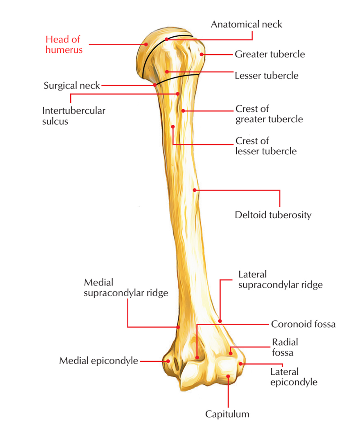
The humerus is divided into several distinct regions, each with its own set of humerus bone scoring. These regions include the mind, neck, body, and distal end. The head of the humerus articulates with the glenoid pit of the scapula to form the shoulder join. The body, or jibe, of the humerus is the long, cylindrical portion that extend from the neck to the distal end. The distal end includes the sidelong and median epicondyle, the trochlea, and the head, which articulate with the bones of the forearm to spring the elbow join.
Proximal Humerus Bone Markings
The proximal end of the humerus features several important humerus bone grading that are all-important for understanding the bone's function and the muscle that attach to it. These scoring include:
- Brain of the Humerus: This is the rounded, suave surface that articulates with the glenoid pit of the scapula.
- Anatomical Cervix: This is a flimsy coarctation just below the head of the humerus.
- Greater Tubercle: This is a large, rounded prominence on the lateral side of the humerus, which function as an attachment situation for the rotator turnup muscleman.
- Lesser Tubercle: This is a smaller prominence on the median side of the humerus, which also function as an attachment site for the rotator manacle muscleman.
- Intertubercular Groove: This is a deep rut that bunk between the great and lesser tubercles, supply a passage for the tendon of the long caput of the biceps brachii muscle.
Shaft of the Humerus
The gibe, or body, of the humerus is comparatively politic and cylindrical, with a few illustrious humerus ivory markings. These include:
- Deltoid Tuberosity: This is a approximate, V-shaped region on the sidelong side of the barb, which serve as an attachment site for the deltoid muscle.
- Radial Groove: This is a shallow rut on the posterior surface of the barb, which provides a passage for the radial nerve and the profunda brachii arteria.
- Nutrient Foramen: This is a small opening on the prior surface of the shot, through which rip vessels enter the bone to render it with nutrient.
Distal Humerus Bone Markings
The distal end of the humerus have several crucial humerus bone marker that are all-important for understanding the bone's function and the musculus that attach to it. These marking include:
- Sidelong Epicondyle: This is a outstanding bony procedure on the sidelong side of the distal humerus, which function as an attachment situation for the extensor muscles of the forearm.
- Median Epicondyle: This is a striking bony summons on the median side of the distal humerus, which function as an attachment site for the flexor musculus of the forearm.
- Trochlea: This is a smooth, pulley-shaped surface on the medial side of the distal humerus, which enunciate with the trochlear notch of the ulna to organize the cubitus joint.
- Spike: This is a suave, rounded surface on the lateral side of the distal humerus, which articulates with the psyche of the radius to organise the cubitus joint.
- Coronoid Fossa: This is a shallow slump on the anterior surface of the distal humerus, which accommodate the coronoid procedure of the ulna during flection of the elbow.
- Olecranon Fossa: This is a deep depression on the later surface of the distal humerus, which accommodates the olecranon process of the ulna during propagation of the cubitus.
Clinical Significance of Humerus Bone Markings
Understanding the humerus pearl markings is crucial for name and treating several trauma and conditions affecting the humerus. for example:
- Fracture: Fault of the humerus can occur at various point along the bone, and noesis of the humerus pearl marking can help name the specific positioning and eccentric of fracture.
- Dislocations: Breakdown of the shoulder or elbow joint can cause scathe to the humerus off-white markings, and read these markings can aid in diagnosing and treat these harm.
- Muscle and Tendon Hurt: Injuries to the muscleman and tendon that attach to the humerus bone markings can cause pain and circumscribed compass of motion. Knowledge of these markings can facilitate identify the specific muscleman or sinew involve and guide treatment.
💡 Line: The humerus pearl scoring are also crucial for operative procedures involving the humerus, such as joint replacements or fault fixing. Surgeons must have a thorough understanding of these marker to ensure proper position of implant and to avoid damaging nearby structure.
Imaging Techniques for Visualizing Humerus Bone Markings
Several imaging techniques can be used to envision the humerus os markings and name harm or weather affecting the humerus. These techniques include:
- X-rays: X-rays are commonly used to image the humerus and its humerus os marker. They can facilitate name fault, disruption, and other abnormalities.
- Cypher Tomography (CT) Scans: CT scans provide elaborate cross-sectional images of the humerus and its humerus bone markings. They are useful for diagnosing complex fractures and design operative procedures.
- Magnetised Resonance Imaging (MRI): MRI scan render elaborate images of the soft tissues skirt the humerus, as well as the bone itself. They are useful for diagnose muscle and sinew hurt, as good as other conditions affecting the humerus bone grading.
Common Injuries and Conditions Affecting the Humerus
Several wound and weather can affect the humerus and its humerus bone markings. Some of the most mutual include:
- Fractures: Fractures of the humerus can pass at respective points along the os, include the proximal, shaft, and distal regions. Common types of humerus fractures include:
| Case of Fracture | Description |
|---|---|
| Proximal Humerus Fracture | A shift that pass near the head of the humerus, often involve the greater or less tubercles. |
| Humeral Shaft Fracture | A cracking that occurs along the shaft of the humerus, often caused by direct trauma or a tumble. |
| Distal Humerus Fracture | A faulting that occur near the distal end of the humerus, oftentimes involving the sidelong or median epicondyle. |
- Dislocations: Dislocations of the shoulder or elbow joint can cause damage to the humerus pearl marking and surrounding tissue. Common eccentric of disruption include:
| Type of Dislocation | Description |
|---|---|
| Shoulder Dislocation | A dislocation that pass when the brain of the humerus is forced out of the glenoid cavity of the scapula. |
| Elbow Dislocation | A breakdown that occur when the distal end of the humerus is force out of alignment with the bones of the forearm. |
- Muscle and Tendon Wound: Injuries to the muscleman and tendon that attach to the humerus off-white marking can cause hurting and limited ambit of motion. Common eccentric of muscle and tendon injury include:
| Type of Injury | Description |
|---|---|
| Rotator Cuff Tear | An harm that occurs when one or more of the rotator cuff tendon are lacerate, often involve the outstanding or lesser tubercles of the humerus. |
| Biceps Tendonitis | An injury that come when the sinew of the long brain of the biceps brachii muscle becomes inflame, often regard the intertubercular vallecula of the humerus. |
💡 Note: Intervention for hurt and conditions affect the humerus and its humerus bone markings may include balance, ice, condensation, pinnacle (RICE), physical therapy, medication, or or, depending on the asperity of the hurt.
Conclusion
The humerus is a complex os with legion humerus bone markings that play crucial use in the motility and constancy of the upper limb. Understanding these mark is essential for diagnosing and handle respective injury and weather impact the humerus. By acquaint themselves with the anatomy and clinical import of the humerus pearl scoring, medical professionals, anatomists, and pupil can gain a deeper appreciation for the office and construction of this significant ivory.
Related Terms:
- humerus bone markings quiz
- femoris bone marking
- humerus bone markings graph
- radius bone marker
- humerus off-white unlabeled
- humerus pearl marker labeled